
 |
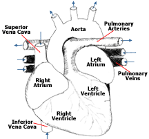
Cardiovascular SystemLike the other mammals, primates have a four-chambered heart and a double- circuit circulatory system and are able to maintain a constant body temperature. The insulating covering is provided by hair, although in the humans nearly all the hair is lost, and insulation is now provided by clothing. The cardiovascular system functions to supply the tissues with oxygen and nutrients by circulating blood throughout the body. At the same time it removes carbon dioxide and other metabolic wastes. The waste products and carbon dioxide move into the blood to be carried away. The process of the circulation of blood is necessary for continued life of the cells, tissues, and ultimately the whole organism. HeartThe heart is made of cardiac muscle, and this is a special type of muscle, because it adjusts the rate of muscular contraction, letting the heart regulate a normal pumping rhythm. The parts of the heart include the chambers, the valves, and the electrical nodes. Heart ChambersThere are two different types of heart chambers. First is the atrium which receives blood that returns to the heart through the veins. The right atruium pumps blood to the right ventricle, and the left atrium pumps blood into the tleft ventricle. The blood is then pumpted from the atrium into the second chamber that is known as the ventricle. The ventricles are larger than the atria and are thicker, muscular walls that are used for aggressively pumping the blood from the heart to the body and lungs. ValvesThe valves which are located within the the heart are placed between the atria and ventricle, and between the ventricles and major arteries. These valves are opened and closed by changes of pressure inside the chambers they function as a barrier to prevent backflow of blood. The vibrations caused by the closing of the respective valves are what create the "lub-dub, lub-dub" heart sounds that are heard through a stethoscope. Electrical NodesThere are two different electrical nodes that are located in the cardiac tissue. The first is the SA, or sinotrial node, which is more commonly called the pacemaker. The pacemaker is located in the wall of the right atrium. It is a small patch of tissue that goes through rhythmic excitation. The impulse rapidly spreads through the atria which causes a muscular contraction and pumping of blood from the atria to the ventricles. The atrioventricular node, or AV, is the other node which relays the impulse of the SA node to the ventricles like a message. The cycle of contraction of heart muscle is also known as the heartbeat. VesselsA vessel is a hollow tube for transportin something. A blood vessel is a hollow tube that transports blood. There are three main types of blood vessels:
The role of these blood vessels is to transpost blood through the entire body and exchange oxygen and nutrients for carbon dioxide and wastes. ArteriesThe arteries are what carry blood away from the heart and are under high pressure because of the the pumping of the heart. In order for them to maintain structure, they have thick, elastic walls to allow stretch and recoil. The large pulmonary artery carries unoxygenated blood from the right ventricles to the lungs where it releases carbon dioxide and receives oxygen. The largest artery though is the aorta. It carries oxygenated blood from the left ventricle to the body. The arteries branch and eventually lead to capillary beds. CapillariesThe capillaries are what make up networks of tiny vessels that have extremely thin, highly permeable walls. They are present in every major tissue of the body and have a role in the exchange of gases, nutrients, and fluids between the blood, body tissues, and alveoli of the lungs. VeinsOn the opposite end of the capillary beds they merge to create veins which return the blood back to the heart. The veins are under a lot less pressure than what the arteries are, and therefore have much thinner walls. The veins contain one-way valves so it may prevent the blood from flowing the wrong direction in the absence of pressure. The pulmonary vein brings back oxygenated blood from the lungs to the left atria. The vena cava returns blood from the body to the right atria. The blood that is returned to the heart is then recycled through the cardiovascular system. |